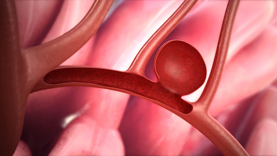
Brain Aneurysm
A brain aneurysm is a weak or thin spot on a blood vessel in the brain that balloons out and fills with blood. Aneurysms can lead to serious health complications, such as stroke or brain damage. It is estimated that up to 6 million people in the United States have an unruptured brain aneurysm, and approximately 30,000 people suffer from a ruptured brain aneurysm (hemorrhage) each year.
More About This Condition
Brain aneurysms can occur when areas of weakened arteries balloon under pressure. Arteries, which carry oxygen-rich blood and nutrients throughout the body, are structured to withstand the pressure caused by normal blood flow. However, they can become damaged as a result of high blood pressure (hypertension), the condition when the force of blood pushing against an artery’s interior wall is consistently too high.
Usually located along major arteries, aneurysms vary in shape and size, sometimes grow over time, and can rupture. Unruptured aneurysms can be problematic because they are susceptible to blood clots forming at the site of the weakened arterial wall. Clot, a mass of thickened blood, can affect the rate of blood flow through a vessel or block it entirely. Also, if a piece of clot breaks off, it can travel through the bloodstream, get stuck in another vessel, and block blood flow at that point. Larger aneurysms can press on nerves or push against the brain.
The sudden rupture of an aneurysm, known as hemorrhagic stroke, leaks blood directly into the brain or into the space between the brain and the skull.
IMPORTANT NOTE: This overview is provided for informational purposes only and should not be used as a substitute for talking with your doctor. Be sure to talk with your doctor for a complete discussion of this condition as well as the benefits and risks of any treatment options.
Symptoms
Unruptured aneurysms may cause symptoms such as blurring of vision and headaches. However, most unruptured aneurysms do not cause symptoms and are discovered by chance during routine brain imaging. Symptoms of ruptured aneurysms can vary depending on the location of the bleed and the amount of brain tissue affected.
Symptoms may include:
- Sudden tingling, weakness, numbness, or paralysis of the face, arm, or leg, particularly on one side of the body
- Sudden, severe headache
- Difficulty with swallowing or vision
- Loss of balance or coordination
- Difficulty understanding, speaking, reading, or writing
- Change in level of consciousness or alertness
Diagnosis
Your doctor may:
- Ask you or your family member about your risk factors, such as high blood pressure, smoking, heart disease, and a personal or family history of stroke.
- Ask about your signs and symptoms and when they began.
- Conduct a physical examination to assess your mental alertness, coordination, and balance.
Your doctor may order one or more of the following tests:
- Computed tomography (CT) scan involves the use of a special X-ray scanner and a computer to create many two-dimensional images, or “slices” of the brain that can be stacked to create a detailed picture. A similar process, called computed tomography angiography (CTA), involves the injection of a special dye, called contrast, via an IV into the bloodstream just prior to scanning. The dye allows doctors to see the blood and blood vessels within the brain tissue.
- Magnetic resonance imaging (MRI) uses computer-generated radio waves and a magnetic field to create two- and three-dimensional detailed images of the brain. Like a CTA, MRA (Magnetic Resonance Angiogram) can provide detailed images of brain vessels but can accomplish this with or without the use of contrast.
- Cerebral angiography involves an injection of contrast dye into the neck and brain arteries. The dye is injected via a catheter, a long tube-like device that is inserted into a leg or arm artery and slowly threaded through the body up to the neck. The dye helps create a detailed image of the arteries on an X-ray in two or three dimensions.
- Cerebrospinal fluid (CSF) analysis measures chemicals in the fluid that cushions the brain and spinal cord. Most often a small amount of cerebrospinal fluid is collected by inserting a thin needle into the spinal cord in the area of the lower back. Additional testing may be needed if test samples indicate bleeding around the brain.
Treatment
Treatment for this condition must always be discussed with your doctor for a full discussion of options, risks, benefits, and other information.
Small, unruptured aneurysms generally do not require treatment unless they grow, trigger symptoms, or rupture. Larger aneurysms may press against a nerve or interfere with blood flow, preventing oxygen and nutrients from reaching other parts of the body. A ruptured aneurysm is considered a medical emergency and immediate treatment may be necessary.
Endovascular embolization is the primary endovascular procedure used to treat both unruptured and ruptured brain aneurysms. A minimally invasive treatment, embolization blocks blood flow to problem areas. To reach an aneurysm, a catheter (tube) is inserted through an incision in the femoral artery at the groin and guided towards the brain. Fluoroscopy (a type of X-ray) is used to track the catheter through the arteries. Once in position, soft platinum metal coils are pushed through the tube and released into the enlarged space. The coils mechanically block the space and induce clotting to cut off blood flow to the affected site. Coils are very small and thin, ranging in size from about twice the width of a human hair (largest) to less than one hair’s width (smallest). The number of coils used depends on the size of the lesion.
Surgical clipping is an open surgical procedure in which blood flow to an aneurysm is cut off by placing a clip at its base.
Learn More
